Surpassing the Human Accuracy: Detecting Gallbladder Cancer from USG Images with Curriculum Learning
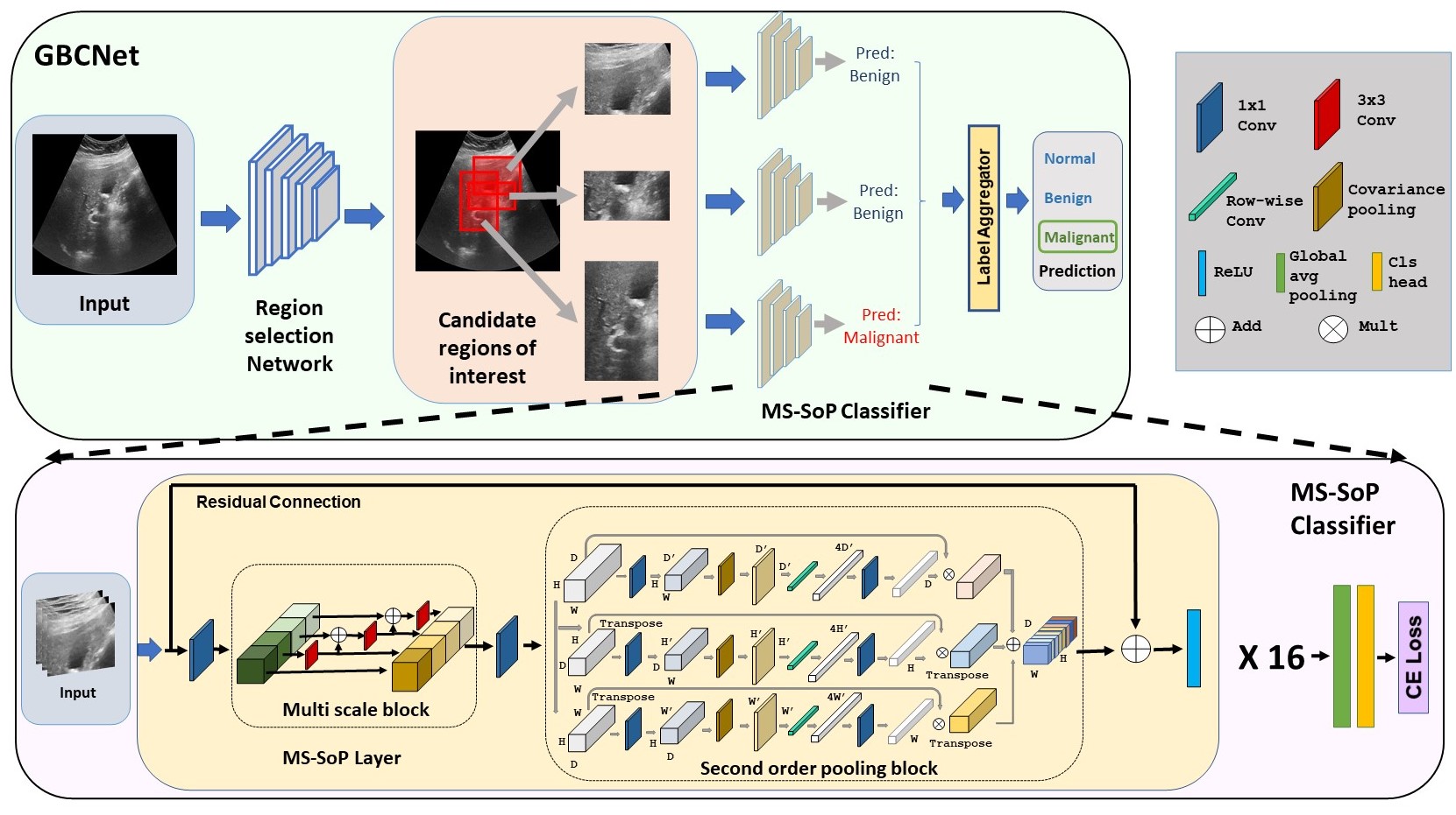
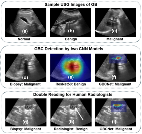
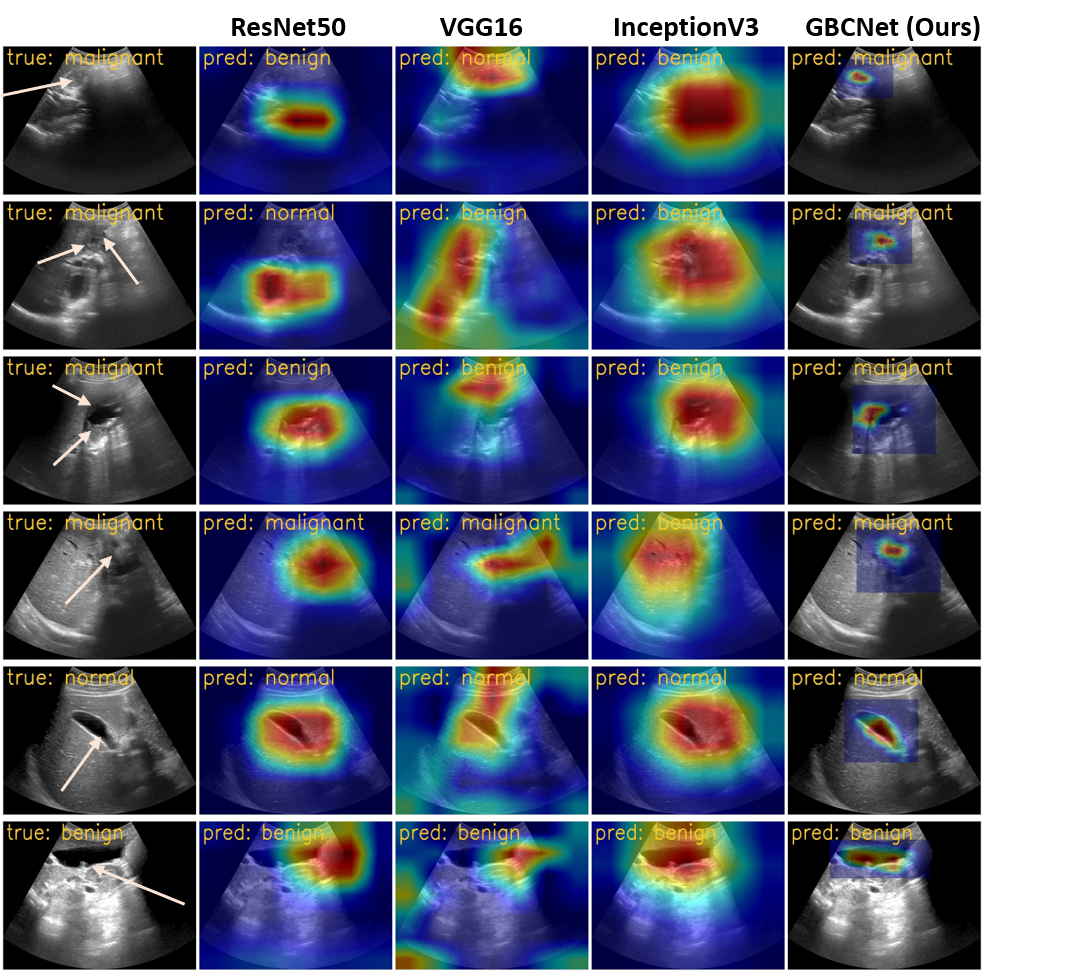
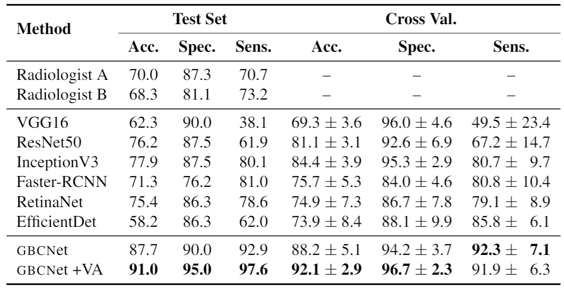
In this work, we explore the potential of CNN-based models for gallbladder cancer (GBC) detection from ultrasound sonography (USG) images as no prior study is known. USG is the most common diagnostic modality for GBC detection due to its low cost and accessibility. However, USG images are challenging to analyze due to low image quality, noise, and varying viewpoints due to the handheld nature of the sensor. Our exhaustive study of state-of-the-art (SOTA) image classification techniques for the problem reveals that they often fail to learn the salient GB region due to the presence of shadows in the USG images. SOTA object detection techniques also achieve low accuracy because of spurious textures due to noise or adjacent organs. We propose GBCNet to tackle the challenges in our problem. GBCNet first extracts the regions of interest (ROIs) by detecting the GB (and not cancer), and then uses a new multi-scale, second-order pooling architecture specializing in classifying GBC. To effectively handle spurious textures, we propose a curriculum inspired by human visual acuity, which reduces the texture biases in GBCNet. Experimental results demonstrate that GBCNet significantly outperforms SOTA CNN models, as well as even the expert radiologists.
Source code and pre-trained models